

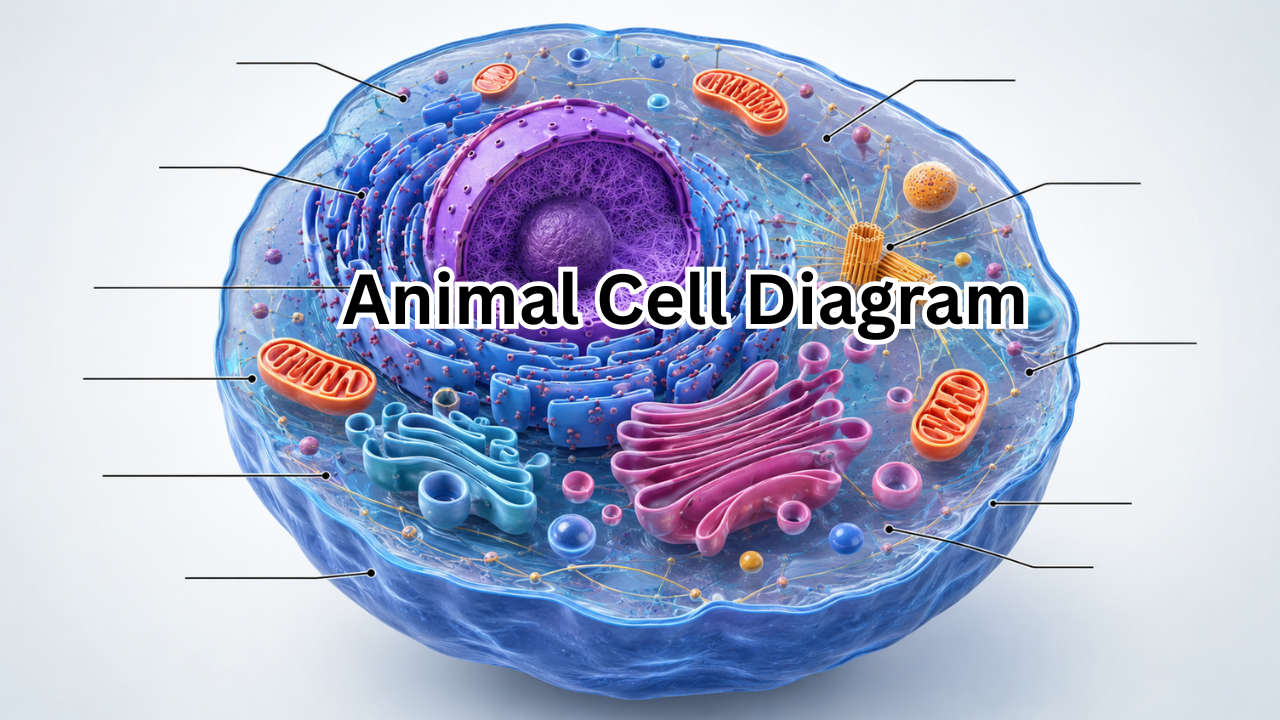
Understanding an animal cell diagram is one of the easiest ways to learn how animal cells are organized and how their different parts work together. Whether you are a school student preparing for a biology exam, a teacher explaining cell structure, or simply curious about basic life science, a clear diagram can make a complicated topic much easier to understand.
An animal cell is the basic structural and functional unit of an animal’s body. Although it is microscopic, it contains several specialized structures called organelles. Each organelle has a particular job, from producing energy and making proteins to storing genetic information and removing waste.
In this guide, we will explore the major parts of an animal cell, explain their functions in simple language, and show how to understand and label an animal cell diagram correctly.
What Is an Animal Cell?
An animal cell is a eukaryotic cell found in animals and other organisms belonging to the animal kingdom. Unlike prokaryotic cells, animal cells contain a true nucleus surrounded by a nuclear membrane.
Animal cells also have several membrane-bound organelles that perform specialized functions. These structures work together to keep the cell alive and allow it to perform essential processes.
One important feature of animal cells is that they do not have a cell wall. They also lack chloroplasts, which are found in plant cells and are responsible for photosynthesis.
Most animal cells are surrounded by a flexible cell membrane, which controls what enters and leaves the cell.
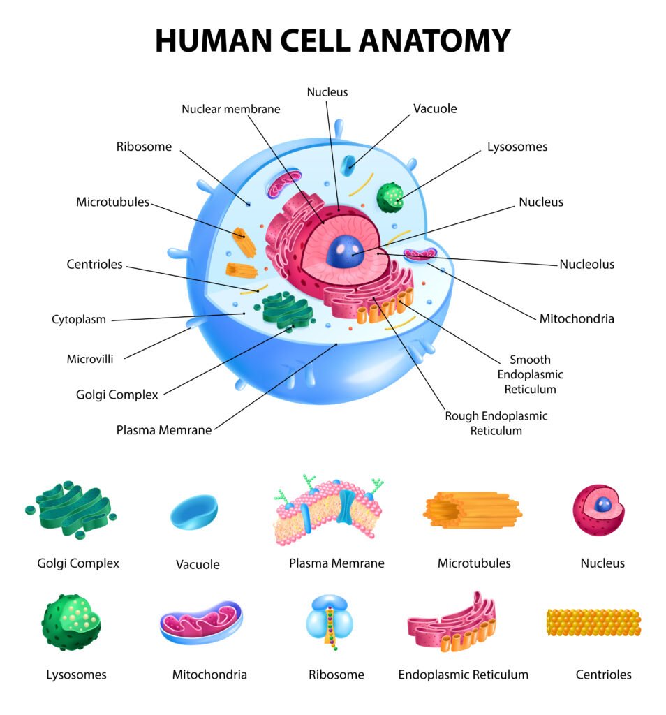
Animal Cell Diagram: Main Parts
A typical animal cell diagram usually shows a rounded or irregularly shaped cell containing several organelles. The exact appearance can vary depending on the type of cell and the diagram style, but the major structures are generally the same.
Here is a quick overview of the most important parts:
| Cell Part | Main Function |
|---|---|
| Cell membrane | Controls movement of substances into and out of the cell |
| Cytoplasm | Provides a medium for many chemical reactions |
| Nucleus | Contains DNA and controls cell activities |
| Nucleolus | Produces components of ribosomes |
| Mitochondria | Produce usable cellular energy |
| Ribosomes | Make proteins |
| Rough endoplasmic reticulum | Produces and transports proteins |
| Smooth endoplasmic reticulum | Helps make lipids and performs other metabolic functions |
| Golgi apparatus | Modifies, sorts, and packages cellular materials |
| Lysosomes | Break down waste and unwanted materials |
| Centrioles | Help organize structures involved in cell division |
| Vacuoles | Store water, nutrients, and other substances |
| Cytoskeleton | Helps maintain cell shape and organization |
Cell Membrane
The cell membrane, also called the plasma membrane, forms the outer boundary of an animal cell.
It is made primarily of a phospholipid bilayer containing proteins and other molecules. Rather than acting as a completely sealed barrier, the membrane is selectively permeable.
This means it controls the movement of substances between the cell and its surroundings.
The cell membrane helps:
- Protect the contents of the cell
- Control the movement of nutrients and waste
- Maintain the cell’s internal environment
- Receive and respond to signals
- Allow communication with neighboring cells
When labeling an animal cell diagram, the cell membrane is usually shown as the thin outer boundary surrounding the entire cell.
Cytoplasm
The cytoplasm is the region inside the cell membrane that contains the organelles.
It consists of the cytosol and the structures suspended within it. Many chemical reactions necessary for cell survival take place in the cytoplasm.
The cytoplasm provides a supportive environment in which organelles can function. It also allows materials to move around inside the cell.
A common mistake is to identify the cytoplasm as simply the space between the cell membrane and nucleus. More accurately, it refers to the cellular material inside the membrane but outside the nucleus.
Nucleus
The nucleus is often one of the most noticeable structures in an animal cell diagram.
It acts as the cell’s main control center because it contains most of the cell’s genetic material, or DNA. DNA provides instructions for producing proteins and regulating many cellular activities.
The nucleus is surrounded by a nuclear envelope, which separates its contents from the cytoplasm.
Important functions of the nucleus include:
- Storing genetic information
- Regulating gene activity
- Helping control cell growth
- Directing protein production
- Participating in cell division
Because of its importance, the nucleus is usually drawn prominently in educational diagrams.
Nucleolus
Inside the nucleus is a dense region called the nucleolus.
The nucleolus is involved mainly in producing ribosomal components. These components eventually form ribosomes, which are essential for protein synthesis.
In a basic animal cell diagram, the nucleolus may appear as a small dark circle within the nucleus.
Mitochondria
Mitochondria are commonly described as the powerhouses of the cell because they generate much of the cell’s usable energy.
They convert energy stored in nutrients into ATP, a molecule cells can use to power many biological processes.
Mitochondria have an outer membrane and a highly folded inner membrane. The folds are called cristae and increase the surface area available for important chemical reactions.
Cells that require large amounts of energy, such as muscle cells, typically contain many mitochondria.
Ribosomes
Ribosomes are tiny structures responsible for protein synthesis.
Proteins are essential for almost every aspect of cellular life. They contribute to cell structure, chemical reactions, communication, transport, and many other processes.
Ribosomes can either float freely in the cytoplasm or attach to the surface of rough endoplasmic reticulum.
In a detailed animal cell diagram, ribosomes may be represented as small dots.
Endoplasmic Reticulum
The endoplasmic reticulum, or ER, is a network of membranes extending through the cell.
There are two major types: rough ER and smooth ER.
Rough Endoplasmic Reticulum
Rough ER has ribosomes attached to its surface, giving it a rough appearance.
Its major functions include producing, folding, and transporting proteins that are destined for particular cellular locations.
It is often shown near the nucleus because the membranes of the rough ER are connected to the nuclear envelope.
Smooth Endoplasmic Reticulum
Smooth ER does not have ribosomes attached to its surface.
It performs several functions, including lipid production, metabolism, and detoxification. In some specialized cells, smooth ER also plays an important role in calcium storage.
Golgi Apparatus
The Golgi apparatus is responsible for modifying, sorting, and packaging proteins and other materials.
You can think of it as a cellular processing and distribution center.
Materials arriving from the endoplasmic reticulum can be modified and packaged into vesicles. These vesicles can then transport their contents to other parts of the cell or toward the cell membrane.
In most animal cell diagrams, the Golgi apparatus looks like a stack of flattened, curved membrane sacs.
Lysosomes
Lysosomes contain enzymes that help break down unwanted materials.
They can digest damaged cellular components, certain foreign materials, and other substances that need to be recycled or removed.
This makes lysosomes important for cellular cleanup and recycling.
They are particularly useful when a cell needs to break down old or damaged organelles.
Centrioles
Centrioles are cylindrical structures associated with the centrosome and are especially important during animal cell division.
They help organize microtubules, which are components of the cytoskeleton. During cell division, this organization contributes to the formation of structures that help separate chromosomes.
In a typical diagram, centrioles may be shown as a small pair of short cylindrical structures near the nucleus.
Vacuoles
Animal cells can contain small vacuoles or vesicles that help store and transport substances.
Unlike plant cells, animal cells generally do not have one large central vacuole. Their storage compartments are usually much smaller.
Depending on the type of animal cell and the diagram being used, these structures may be shown as small membrane-bound sacs.
Cytoskeleton
The cytoskeleton is a network of protein fibers that helps maintain the cell’s shape and organization.
It is not a rigid skeleton like the bones in an animal body. Instead, it is a dynamic internal framework.
The cytoskeleton helps with:
- Maintaining cell shape
- Moving organelles
- Transporting materials inside the cell
- Cell movement
- Cell division
- Organizing internal structures
Some simplified animal cell diagrams do not label the cytoskeleton, but detailed diagrams may include it.
How to Read an Animal Cell Diagram
Looking at a diagram with many labels can initially feel confusing. A useful strategy is to identify structures based on their position and appearance.
Start with the outermost boundary. This is usually the cell membrane.
Next, locate the nucleus. It is often one of the largest structures inside the cell and may contain a visible nucleolus.
Then look for the mitochondria, which are commonly drawn as oval or bean-shaped structures with internal folds.
The rough ER is generally shown as a membrane network covered with small dots representing ribosomes. The Golgi apparatus usually appears as a stack of curved flattened sacs.
This approach makes it easier to understand an unlabeled animal cell diagram.
Animal Cell vs. Plant Cell
Animal and plant cells share many structures because both are eukaryotic cells. However, they also have several important differences.
| Feature | Animal Cell | Plant Cell |
| Cell membrane | Present | Present |
| Cell wall | Absent | Present |
| Nucleus | Present | Present |
| Mitochondria | Present | Present |
| Chloroplasts | Absent | Present |
| Large central vacuole | Usually absent | Usually present |
| Typical shape | Flexible and varied | Often more regular |
| Centrioles | Commonly present | Generally absent in higher plants |
These differences are useful when studying an animal cell diagram alongside a plant cell diagram.
One of the easiest ways to distinguish them is to look for a cell wall and chloroplasts. If those structures are clearly present, the diagram is likely showing a plant cell rather than a typical animal cell.
Why Animal Cell Diagrams Are Important
Diagrams are particularly useful in biology because cells are too small to observe clearly with the naked eye.
A well-labeled diagram turns an invisible microscopic structure into something students can study visually.
Animal cell diagrams can help students:
- Memorize organelles
- Understand relationships between cell structures
- Prepare for biology exams
- Learn organelle functions
- Compare animal and plant cells
- Practice biological labeling
Teachers can also use diagrams to explain how different organelles cooperate rather than treating each organelle as an isolated structure.
Easy Way to Memorize Cell Organelles
Instead of memorizing a long list of names and definitions at once, group the organelles according to their general jobs.
Control and information
- Nucleus
- Nucleolus
Energy
- Mitochondria
Protein production
- Ribosomes
- Rough ER
Processing and transportation
- Smooth ER
- Golgi apparatus
- Vesicles
Cleaning and recycling
- Lysosomes
Structure and movement
- Cytoskeleton
- Centrioles
Protection and exchange
- Cell membrane
This method can make revision much easier because you are learning how the parts relate to their functions.
Common Mistakes When Labeling an Animal Cell
Students often lose marks not because they do not know the organelles, but because they confuse similar-looking structures.
Some common mistakes include:
- Calling the cell membrane a cell wall
- Confusing rough ER with smooth ER
- Forgetting that ribosomes are responsible for protein synthesis
- Identifying the Golgi apparatus as the endoplasmic reticulum
- Assuming animal cells contain chloroplasts
- Drawing one large central vacuole like a typical plant cell
- Mixing up the nucleus and nucleolus
A good way to avoid these errors is to learn both the appearance and function of each structure.
Tips for Drawing an Animal Cell Diagram
If you need to draw an animal cell for a school assignment or examination, keep the diagram clear rather than making it unnecessarily complicated.
A few useful tips are:
- Draw a large, rounded cell outline to represent the cell membrane.
- Add the nucleus as a prominent structure inside the cell.
- Include mitochondria with their characteristic internal folds.
- Add rough and smooth ER near the nucleus.
- Draw the Golgi apparatus as stacked curved membranes.
- Include smaller structures such as lysosomes and ribosomes.
- Add centrioles if required by your syllabus.
- Use straight labeling lines that do not cross each other.
- Keep labels readable and correctly spelled.
- Add a title such as “Animal Cell” above the diagram.
The level of detail should match the assignment. A simple school diagram does not need to look like a professional scientific illustration.
Frequently Asked Questions About Animal Cell Diagrams
What is an animal cell diagram?
An animal cell diagram is a visual representation of an animal cell that identifies its major structures and organelles. It is commonly used in biology education to explain cell organization and the functions of different parts.
What are the main parts of an animal cell?
The major parts commonly shown include the cell membrane, cytoplasm, nucleus, mitochondria, ribosomes, endoplasmic reticulum, Golgi apparatus, lysosomes, and centrioles. More detailed diagrams may include additional structures.
Does an animal cell have a cell wall?
No. Typical animal cells do not have a cell wall. They are surrounded by a flexible cell membrane. This is one of the major differences between animal and plant cells.
Do animal cells have chloroplasts?
No. Animal cells do not normally contain chloroplasts because they do not perform photosynthesis. Chloroplasts are characteristic of plant cells and certain other photosynthetic organisms.
What is the function of the nucleus in an animal cell?
The nucleus stores most of the cell’s DNA and helps regulate gene activity and many cellular processes. It plays a central role in controlling cell functions and cell reproduction.
Why are mitochondria called the powerhouse of the cell?
Mitochondria produce ATP through cellular respiration, providing energy that cells can use for many activities. Because of this major role in energy production, they are commonly called the powerhouse of the cell.
Final Thoughts
Learning an animal cell diagram becomes much easier when you stop treating it as a collection of unfamiliar labels and start seeing it as a coordinated system. Every organelle has a role, and those roles are connected.
The nucleus stores genetic instructions, ribosomes help build proteins, mitochondria provide usable energy, the endoplasmic reticulum helps produce and transport materials, and the Golgi apparatus processes and distributes them. Meanwhile, lysosomes help with cellular cleanup, while the cell membrane controls interaction with the outside environment.
Once you understand these basic relationships, labeling an animal cell diagram becomes far less difficult. Whether you are studying for an exam, preparing a classroom presentation, or reviewing basic biology, understanding the structure and function of each organelle will give you a much stronger foundation for more advanced topics in life science.
Also Read : Houzz Pro: A Complete Guide to Features, Pricing, Benefits, and How It Works










Leave a Reply